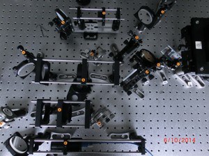
Surface Plasmon Resonance (SPR) Microscope
In our research we focus on interfaces between electronic chips or electrodes and animal cells.
With the surface plasmon resonance microscope (SPR), we can monitor the interface between chips and neurons in real time (in the imaging mode) and measure the distance between the cell membrane and the gold surface (in the scanning mode). The measurements work in situ and without chemical labeling.
The samples consist of sapphire chips coated with a gold layer incorporated in petridishs. Cells are cultured on top of the gold surface. For the life imaging, we use the imaging mode of the SPR microscope. Illuminating the sample from the bottom with an objective, the laser light excites surface plasmons - collective oscillations of electrons. Their energy depends on the surrounding of the gold surface.
A fraction of the incident light which matches the excitation condition of the plasmons, is absorbed whereas the rest is reflected. The reflected light is recorded by a CMOS camera and shows the shape of the cells in the x-y- plane.
In a scanning mode, the incident light in focused tightly. The reflected light is recorded by a second CMOS-camera. From the reflected spectrum we extract information about the excited plasmons. This can be mapped on the distance between the chip surface and the membrane. Thus we can measure distances of several 10 nanometers in z-direction.
| Some characteristic data: | |
| Laser | 10 mW HeNe laser (λ = 632.8 nm, Melles Griot) |
| Objective | 100x 1.65 numerical aperture oil objective (OBJ, Olympus) |
| Camera | CMOS cameras (CMOS, XIMEA) |
Contact:
Justus Bednar
Tel.: +49-2461-61-2331
e-mail: ju.bednar@fz-juelich.de
Prof. Dr. Andreas Offenhäusser
Tel.: +49-2461-61-2330
e-mail: a.offenhaeusser@fz-juelich.de