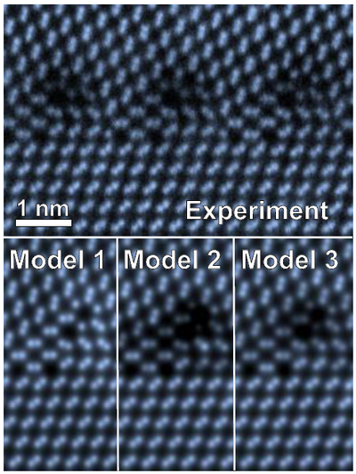
Quantitative modelling
Transmission electron microscopy (TEM) is a versatile technique that provides images in which the contrast is strongly influenced by the material structure at the atomic level. In most cases, it is very challenging to go beyond qualitative statements based on electron microscopic images and to quantitatively determine material properties. The main reasons for this complication are the strong interaction of electrons with matter via the Coulomb force, a variety of interaction phenomena leading to a multitude of signal dependencies, and the quantum mechanical nature of electron scattering and electron optical imaging. Our goal is to use and develop the modelling of electron scattering and imaging to the point of achieving near perfect agreement between experiment and simulation. The example below compares an experimental image to images calculated from atomic structure models taking into account the detailed knowledge of our electron microscopes. The results of atomic-scale structural modelling in TEM can also provide reality checks to accompany the calculation of material properties by molecular dynamics or density functional theory, or even provide initial values for such approaches.
Elastic electron scattering and scattering due to the excitation of phonons can be modelled by means of software packages such as the Dr. Probe software [2] which is developed at the Ernst Ruska-Centre. We continuously work on extending image simulation software to include experimental techniques that go beyond the usual routines with the goal of making them available to a broader community. Among such techniques are electron energy loss spectroscopy and secondary X-ray emissions. They provide a signal from the chemical composition of a material via the spectral analysis of inelastic electron scattering. The fine structures of electron energy-loss spectra also contain information about chemical bonding and magnetic sample properties. Analysing the fine structure of a spectrum is extremely challenging, especially because a very weak signal often needs to be separated from a strong background and may additionally be affected by multiple scattering. The latter is very often due to a larger sample thickness used in such experiments in order to improve signal quality. Modelling the multitude of phenomena of electron scattering in a material helps on the one hand to provide insights into the relations between material properties and signal recorded in the experiment, and on the other hand to advance the design of experimental setups and new instruments with respect to improving the signal quality [3,4].
Literature:
[1] A. Stoffers, B. Ziebarth, J. Barthel, O. Cojocaru-Mirédin, C. Elsässer, D. Raabe, Phys. Rev. Lett 115 (2015) 235502 (doi: 10.1103/PhysRevLett.115.235502)
[2] J. Barthel, DrProbe - A software for high-resolution STEM image simulation, Ultramicroscopy 193 (2018) 1-11 (doi: 10.1016/j.ultramic.2018.06.003)
[3] J. Barthel, J. Mayer, J. Rusz, P.-L. Ho, X. Y. Zhong, M. Lentzen, R. E. Dunin-Borkowski, K. W. Urban, H. G. Brown, S. D. Findlay, and L. J. Allen, Understanding electron magnetic circular dichroism in a transition potential approach, Phys. Rev. B 97 (2018) 144103 (doi: 10.1103/PhysRevB.97.144103)
[4] J. Barthel, M. Cattaneo, B.G. Mendis, S.D. Findlay, L.J. Allen, Angular dependence of fast-electron scattering from materials, Phys. Rev. B 101 (2020) 184109 (doi: 10.1103/PhysRevB.101.184109)
Contact:
