Using whole-brain laminar fMRI to investigate new dimensions of the human neocortex
It is known that resting-state networks in the brain sustain conscious awareness and diverse cognitive processing in the absence of tasks and can provide information relating to the functional architecture of the brain. However, although resting-state networks in cortical areas of the brain have been studied in relative detail, the cortical depth across different networks has not yet been extensively investigated. This has been mainly due to the small brain coverage enforced in high-resolution imaging methods.
This research, published in Frontiers in Neuroscience, builds on a novel fMRI technique developed by researchers at the Forschungszentrum Jülich that combines echo-planar-imaging with keyhole (EPIK) with repetition-time-external (TR-external) EPI phase correction to provide a spatial resolution as high as half-millimetre in-plane (0.51 × 0.51 × 1.00 mm3 (0.26 mm3 voxel)) or 0.63 mm isotropic (i.e. in all voxel dimensions) with whole-cerebrum coverage.
Dr Seong Dae Yun, the physicist behind the sequence technical improvements and first author of the publication detailing the recent version of the sequence (Mapping of Whole-Brain Resting-State Networks with Half-Millimetre Resolution), commented, “We have been working on the development of the sequence for years. Bringing the voxel volume down to 0.25mm3, finally possible at high field (7T), has been very challenging because, theoretically, we could keep on reducing the voxel size, but then the amount of signal coming from each voxel would be too low to distinguish real neural activity from noise, so we are approaching a point where the intrinsic limitations of MRI need to be considered… still, this improvement in coverage and resolution is a great milestone because it opens the possibility of analysing the function of structures that could not be distinguished from each other due to their small size, without losing the field of view (e.g. different cortical depth territories across the whole cerebrum)”.
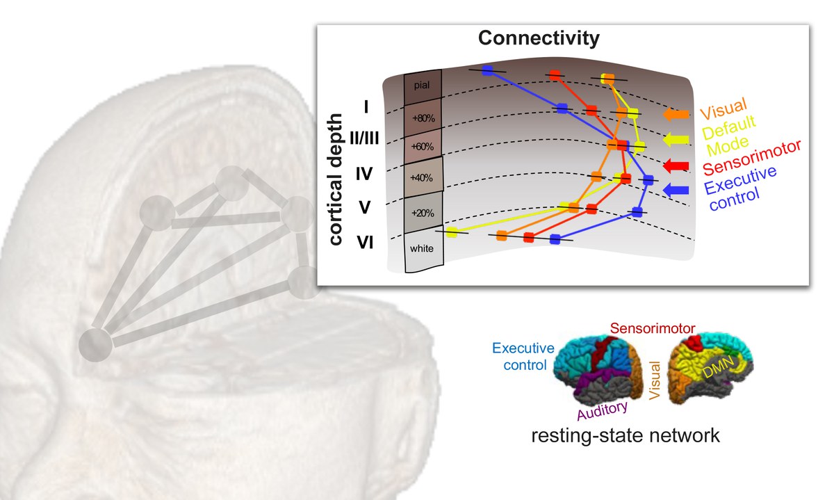
Based on fMRI data from 13 healthy volunteers, the newly developed sequence was used to demonstrate the dependence of network connectivity on the cortical depth; for instance, the involvement of the supragranular layers of the cerebral cortex appears to be critical in the maintenance of resting-state dynamics within the ‘default mode network’ (a group of areas that are more active when subjects are asked to remain awake but without thinking about anything in particular – usually related to mind-wandering). Given the cytoarchitectonics of the human neocortex, and based on the results obtained, the cortical thickness constitutes an important dimension in the characterisation of resting-state oscillations in the healthy brain, and its functional study may facilitate the identification of targets in neurological diseases from a novel perspective.
Lead author Dr Patricia Pais-Roldán said, “I feel honoured to be working within this team where cutting-edge sequences are being developed. This atmosphere fosters novel research that cannot be conducted in most labs. Studying the function of the cortex with such detail (along its depth, while preserving the near whole-brain mapping) in living human subjects is bringing unforeseen results. We are starting to use the method to detect potential differences between diverse mental states, and I am convinced this line of research will help in deciphering novel aspects of psychological disorders”.
Origional publication:
Cortical depth-dependent human fMRI of resting-state networks using EPIK