Polarization Microscopy
I stumbled upon the idea of formalin fixed tissue much later and realized with satisfaction, that formalin fixation does not impair the birefringence of myelinated nerve fibers. Therefore, we can study nerve fibers hardened and conserved in formalin … with polarization microscopy and observe possible pathological alterations
(K. Brodmann, J Psych Neurol, 1903).
We have brought this observation into the 21st century, developing and adapting polarimetric technologies to the requirements of high-throughput large-scale polarization microscopy of hair-thin, unstained brain sections. We have named the approach 3D Polarized Light Imaging (3D-PLI), since we are aiming for contrasting distinct fibers and tracts and investigating their three-dimensional courses across serial brain sections.
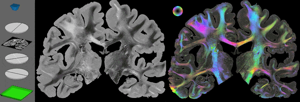
High quality polarimetric measurements require specific preparation and handling of the studied brain tissue in order to preserve the integrity of the myelin sheaths which are responsible for the optical effect referred to as birefringence. Cryo-sectioning of formalin conserved and glycerol soaked tissue was demonstrated to comply with this condition. During sectioning, en face images of the remaining brain block are acquired (blockface imaging) serving as reference images for later reconstruction of the initial brain shape. The generated unstained histological brain sections are scanned with different types of polarimetric setups, which are generally speaking composed of two rotating linear polarizers, one quarter-wave retarder and a green-wavelength light source (see above figure, left). High-performance computing-based signal and image analysis as well as simulation approaches finally enable reliable interpretation and visualization of the targeted fiber architecture (see above figure, right).